The Difference Between Male And Female Pelvis
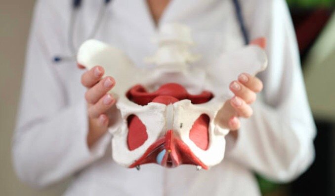
- The female pelvis consists of a wider sacrum and a wider subpubic angle when compared to males.
- The female pelvis’ ischial spines are also less prominent than the male’s ischial spines.
- The male pelvis’ sacrum is generally longer and more curved with a narrower sub-pubic arch. In females a wider pelvic aperture is needed as it functions as the birth canal during labour.
But our major focus of today is the female Pelvis.
THE MEANING OF FEMALE PELVIS

There are some structural differences between the female and the male pelvis.
- Most of these differences involve providing enough space for a baby to develop and pass through the birth canal of the female pelvis.
- As a result, the female pelvis is generally broader and wider than the male pelvis.
FEMALE PELVIS. BONES
There are two hip bones, one on the left side of the body and the other on the right.
- Together, they form the part of the pelvis called the pelvic girdle.
- The hip bones join to the upper part of the skeleton through attachment at the sacrum.
- Each hip bone is made of three smaller bones that fuse together during adolescence:
These bones are:
- Ilium: The largest part of the hip bone; ilium is broad and fan-shaped.
- Pubis: The pubis bone of each hip bone connects to the other at a joint called the pubis symphysis.
- Ischium: this is paired bone of the pelvis ( combination of the ilium and pubis)
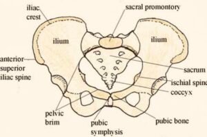
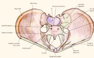
- The innominate bones are joined anteriorly at the symphysis pubis and each articulates posteriorly with the sacrum in the sacroiliac joints. All three joints are fixed in the non-pregnant state, but during pregnancy there is relaxation of the joints to allow some mobility during labour and birth. The sacrum articulates with the fifth lumbar vertebra superiorly and the coccyx inferiorly.
- The bony pelvis is divided into the
- false pelvis and the
- true pelvis by the pelvic brim.
- The true pelvis is divided into three sections: the pelvic inlet (bounded anteriorly by the superior surface of the pubic bones and posteriorly by the promontory and alae of the sacrum); the mid-pelvis (at the level of the ischial spines); and the pelvic outlet (bounded anteriorly by the lower border of the symphysis, laterally by the ischial tuberosities and posteriorly by the tip of the sacrum).
THE EXTERNAL GENITALIA
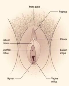
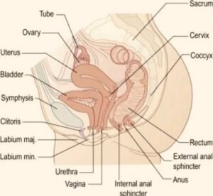
The term vulva is generally used to describe the female external genitalia, and includes the mons pubis, the labia majora, the labia minora, the clitoris, the external urinary meatus, the vestibule of the vagina, the vaginal orifice and the hymen.
THE INTERNAL GENITALIA
The internal genitalia include the vagina, the uterus, the Fallopian tubes and the ovaries.
- Situated in the pelvic cavity, these structures lie in close proximity to the urethra and urinary bladder anteriorly and the rectum, anal canal and pelvic colon posteriorly.
TYPES OF PELVIS
In midwifery, the shape of the pelvic cradle is an important component in determining the outcome of the birth experience.
Basically there are 4 types of pelvis
- And these are classified according to the shape of the brim or inlet.
- Although the shape of the pelvis varies, it is not a rigid, fixed structure, but an elastic system of bones that can widen and stretch, and which is very flexible at the joints so that it can open wide during labor.
- The mother’s mobility and position during labor are critical in order to facilitate this process and there is a lot to be said for active labor if a mother does not have what is considered the ideal shape pelvis for birth to happen easily.
Types of Pelvis are:
- Android pelvis
- Anthropoid pelvis
- Gynaecoid (genuine female Pelvis )
- Platypelloid pelvis
1. THE ANDROID PELVIS
- It has a heart-shaped brim and it is quite narrow in front.
- This type of pelvis is likely to occur in tall women with narrow hips and it is also found in African women.
- The pelvic cavity and outlet is often narrow, straight and long.
- The ischial spines are prominent.
- Women with this shape pelvis may have babies that lie with their backs against their mothers’ backs and may experience longer labours.
- It is important that these women take an active role during their labour and need to squat and move around as much as possible.
2. THE ANTHROPOID PELVIS
- It has an oval brim and a slightly narrow pelvic cavity.
- The outlet is large, although some of the other diameters may be reduced.
- If the baby engages in the pelvis in an anterior position, labour would be expected to be straightforward in most cases.
3. THE GYNAECOID OR GENUINE FEMALE PELVIS
- It has an almost round brim and will permit the passage of an average-sized baby with the least amount of trauma to the mother and baby in normal circumstances.
- The pelvic cavity (the inside of the pelvis) is usually shallow, with straight side walls and with the ischial spines not so prominent as to cause a problem as the baby moves through.
4. THE PLATYPELLOID PELVIS
- It has a kidney-shaped brim and the pelvic cavity is usually shallow and may be narrow in the anteroposterior (front to back) diameter.
- The outlet is usually roomy.
- During labour, the baby may have difficulty entering the pelvis, but once in, there should be no further difficulty.
- Many women are concerned that their pelvic capacity may be limited and that they will therefore have difficulty in giving birth.
- The true capacity of the pelvis will only be realized during labor by the midwife. Only when the forces created by the mother and baby during birth will the pelvis open to its full potential. This may take some time, but it is the only true way of exploring the “fit” between the mother and baby during birth.
CLASSIFICATION OF PELVIS BY CALDWELL MOLOY AND THEIR DIAMETERS (1933)
Caldwell- Moloy Classification of Pelvic Types (1933).

Four types of female pelves were described. Actually, the majority of pelves are of mixed types:
- Gynaecoid pelvis(50%):
- It is the normal female type.
- Inlet is slightly transverse oval.
- sacrum is wide with average concavity and inclination.
- Side walls are straight with blunt ischial spines.
- Sacro-sciatic notch is wide.
- Subpubic angle is 90-100⁰.
- Anthropoid pelvis (25%):
- It is ape-like type.
- All anteroposterior diameters are long.
- All transverse diameters are short.
- Sacrum is long and narrow.
- Sacro-sciatic notch is wide.
- Subpubic angle is narrow.
- Android pelvis (20%):
- it is a male type.
- Inlet is triangular or heart-shaped with anterior narrow apex.
- Side walls are converging (funnel pelvis) with projecting ischial spines.
- Sacro-sciatic notch is narrow.
- Subpubic angle is narrow 90⁰.
- Platypelloid pelvis (5%):
- It is a flat female type.
- All anteroposterior diameters are short.
- All transverse diameters are long.
- Sacro-sciatic notch is narrow.
Subpubic angle is wide.
DIAMETERS OF THE PELVIS
Diameters of the Pelvis are classified into three (3).
These are:
- Anterior
- Transverse
- Oblique
The anterior-posterior diameter is the line from the sacral promontory to the upper border of the symphysis pubis. It is measured up to 11cm.
The transverse diameter or lateral is the distance between the farthest points on the pelvic brim over the illiopectineal lines. It measures up to 13cm. It is the largest diameter of the pelvis (Hence option 2 is correct).
Oblique diameter is measured up to 12cm and is the line from the sacroiliac joint to the opposite illiopectineal Eminence.
Please take note of these diameters for exam purposes.
However,
“A Female pelvis is described as a skeletal ring formed by two innominate bones and sacrum and coccyx.
NOTE:
- The pelvis is the bony canal through which the fetus passes during the birth of the baby.
- It comprises three parts: Brim; Cavity and Outlet.
- The Brim is formed by the sacrum posteriorly, the iliac bones laterally, and the pubic bone from the anterior.
LIGAMENTS OF THE PELVIS
The bony integrity of the pelvis is supported by a variety of ligaments that lend crucial flexible strength to the pelvic cavity and support some of the internal pelvic structures.
- The principal ligaments are the sacrotuberous, sacrospinous, and iliolumbar, and in the female pelvis, there are further ligaments to support the ovaries and uterus
- Trauma or stretching of these ligaments can result in pain syndromes, causing a significant impact on quality of life if not recognized and treated appropriately.
- Furthermore, pelvic floor dysfunction, which describes wider pathology involving the entire pelvic floor musculature and ligaments, can have a deleterious effect on urinary, bowel, and sexual function.
STRUCTURE AND FUNCTION OF THE FEMALE PELVIS
The key ligaments of the pelvis present in both sexes are the
- sacrotuberous
- sacrospinous, and
- iliolumbar ligaments.
Further ligaments include the
- anterior sacroiliac,
- anterior
- sacrococcygeal,
- posterior sacroiliac, posterior
- sacrococcygeal, and
- pectineal ligaments.
Anterior Articulations, Pelvis, and Posterior articulations, Pelvis. Further specific female pelvic ligaments are the broad ligament and ligaments of the ovaries and uterus.
LIGAMENTS OF THE PELVIS
Pelvic ligaments can be categorised according to their associated joints and are therefore divided into the ligaments of the
- Lumbosacral
- Sacroiliac
- Sacrococcygeal
- Pubic symphyseal
1. LUMBOSACRAL LIGAMENTS (ILIOLUMBAR LIGAMENTS)
- The iliolumbar ligament is composed of thick and strong fibrous bands of connective tissue originating from the tip of the transverse process of the fifth lumbar vertebra.
- Here, it extends laterally as a superior and inferior band, resembling a V-shape.
- The superior band inserts at the iliac crest, and the inferior band blends with the front of the anterior sacroiliac ligament on the pelvic surface of the ileum.
- This ligament stabilizes and strengthens the lumbosacral joint, restricting the rotational movement at the lumbosacral joint.
2. SACROILIAC LIGAMENTS
- The sacroiliac joint complex is reinforced by various ligaments that provide structural support to this important joint within the pelvis.
- The interosseous sacroiliac ligament provides the largest and most robust support of the sacroiliac joint in both females and males.
- It attaches at deep pits on the lateral mass of the posterior surface of the sacrum, extending to the ilium n a series of short and very strong fibers.
- As mentioned above, its anterosuperior surface blends with the iliolumbar ligament. Furthermore, the most superficial fibers of the posterior aspect of the interosseous sacroiliac ligament form the posterior sacroiliac ligament.
- The anterior sacroiliac ligament is a band of connective tissue that connects the anterior surface of the lateral part of the sacrum to the margin of the auricular surface of the ilium.
3. SACROCOCCYGEAL LIGAMENTS
- The sacrococcygeal joint is reinforced with anterior, posterior, and lateral ligaments.
- The anterior sacrococcygeal ligament unifies the anteriormost aspect of the respective bone bones.
- Posteriorly there are two posterior sacrococcygeal ligaments that perform a similar function on the dorsal aspect of the joint.
- Finally, two lateral sacrococcygeal ligaments run from the coccygeal transverse processes inferiorly to the angle of the sacrum.
4. PUBIC SYMPHYSEAL ( LIGAMENTS OF THE PUBIS SYMPHYSIS).
- The pubic symphysis is a midline cartilaginous joint uniting the two pubic bones. Small ligamentous supports to this joint are present in the form of the superior pubic ligament and the arcuate pubic ligament inferiorly.
- The strength and contribution to joint stability provided by these ligaments are controversial.
- However, it is accepted that the arcuate pubic ligament is stronger than its superior counterpart.
OTHER LIGAMENTS OF THE PELVIS INCLUDE
5. SACROTUBEROUS LIGAMENTS
- The sacrotuberous ligament is a fan-shaped fibrous band of connective tissue with great strength, providing stability to the posterior pelvis.
- Its wider superior origin runs from the transverse tubercles of the sacrum below the level of the sacroiliac joint, the posterior superior and posterior inferior iliac spines, and the upper coccyx, downwards to attach to the tuberosity of the ischial tuberosity.
- The average length of the sacrotuberous ligament from cadaveric dissections is 70 mm in women and 64 mm in men.
- The sacrotuberous ligament is blended with the posterior sacroiliac ligament, which is a continuation of the superficial fibers of the sacroiliac ligament, which attaches the sacrum firmly to the ilium.
6. THE SACROSPINOUS LIGAMENTS
- The Sacrospinous ligament is a triangular band of connective tissue that lies on the pelvic aspect of the sacrotuberous ligament.
- It has a broad base with origins at the lower sacrum and upper coccyx.
- It travels laterally from its base, narrowing as it does so, to attach to the spine of the ischium and has an average length of 46 mm in women and 38 mm in men.
- The sacrospinous ligament divides the greater sciatic notch to the greater sciatic foramen and the lesser sciatic foramen, through with pass several important neurovascular structures critical to the lower limb and genital function, including the gluteal, pudendal, and sciatic nerves and the gluteal and pudendal vessels.
THE FEMALE PELVIS LIGAMENT
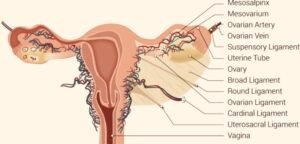
- The broad ligament is a sheet of pelvic peritoneum extending bilaterally from the lateral pelvic sidewalls to the uterus in the midline.
- The broad ligament folds over the fallopian tubes and ovaries and covers them anteriorly and posteriorly and is subdivided into the
- mesovarium,
- mesosalpinx, and
- mesometrium.
- 1. The mesovarium is the part of the broad ligament related to the ovaries.
- This posterior sheet of broad ligament extends to the hilum of the ovary and contains the neurovascular tissue of the ovary, here as the suspensory ligament of the ovary.
- The peritoneal fold of the broad ligament does not cover the surface of the ovary, but rather the anterior border of the ovary is attached to the broad ligament.
- The mesosalpinx is the part of the broad ligament related to the fallopian tube.
- Here, the broad ligament folds over and encloses the fallopian tube.
- The mesometrium is the part of the broad ligament related to the uterus.
- From the uterus, the broad ligaments fan out laterally while covering the external iliac vessels.
- The mesometrium is the largest section of the broad ligament.
Uterine Tubal Anatomy and Ligaments. Anatomical structures surrounding the uterus and fallopian or uterine tubes, including the mesosalpinx, mesovarium, ovarian artery, ovarian vein, suspensory ligament, uterine tube, ovary, broad ligament, round ligament, ovarian ligament, cardinal ligament, uterosacral ligament, and vagina. Contributed by B Palmer.
