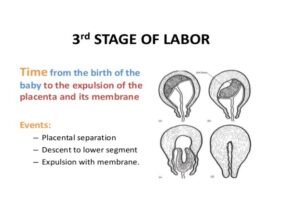
The Mechanical Factors
The uterus reduces in size 2.5cm below the umbilicus, or 15cm above the symphysis pubis after the expulsion of the fetus. The contraction and retraction of the uterine muscles continues. The placental site is reduced to half. Since the placenta is inelastic, it does not contract, so it detaches from the shrinking uterine wall. The placenta is pushed further to the lower.
This is accumulated blood from the separated placenta. With the next contraction the placenta is pushed into the vagina and expelled.
You will know that the placenta has separated and has been expelled from the upper uterine segment into the lower segment or into the vagina when you have noted:
Elongation of the cord which does not recede on pressing at the symphysis pubis
A gush of blood.
The uterus contracts like a cricket ball.
The placenta is expelled either with maternal side exposed, known as the Mathew-Duncan method or the foetal side exposed known as the Shultz Method.
Control of Bleeding After Delivery
Steps should be taken to ensure the control of bleeding:
- The uterine muscle’s contraction and retraction causes the placental site to reduce into half. Criss-cross fibres control bleeding by compressing the blood vessels. These fibresare also known as ‘living ligatures’
- Clotting of blood takes place in the sinuses sealing the bleeding points a few hours later when uterine contractions are less vigorous.
- The time interval between the delivery of the baby and delivery of the placenta is a dangerous period, in which one of the greatest complications of pregnancy and labour can occur.
- This complication is excessive bleeding or postpartum haemorrhage (PPH). You should never leave the mother alone even for a short while during this stage.
- The third stage of labour can be managed either passively or actively.
The Passive or Natural Method of Managing the Third Stage of Labour
The passive or natural method occurs naturally, that is without any interference. For example, in a normal delivery, if oxytoxic drugs are not used, the uterus generally remains inactive for a few minutes after the delivery of the baby, after which regular contractions then begin again.
Physiology of the third stage takes place, the placenta is expelled and bleeding is controlled.
Giving Oxytocic Drugs
Oxytocin, ergometrine or syntometrine stimulate uterine contraction.
Ergometrine 0.5mg given IM causes a uterine contraction to occur five to seven minutes after the injection. Given intravenously it acts within 45 seconds.
Syntometrine is a mixture of oxytocin and ergometrine 0.5ml given IM acts within two to three minutes.
Usually sytrometrine is given with the delivery of the anterior shoulder while ergometrine is given at the crowning of the head.
Controlled Cord Traction
The uterus must be firmly contracted before this method can be used. It is not necessary to wait for signs of placenta separation.
Place the left hand above the symphyis pubis, push the uterus upwards and backwards with the right hand and pull the cord downwards and outwards.
Apply traction steadily without jerking When the placenta is visible at the vulva, direct the cord upwards.
The placenta will follow the curve of the birth canal. Receive it with both hands and rotate the placenta. This will twist the membranes. You can then deliver the membranes with up side movements, which enable them to be drawn out without breaking.
Maternal Effort
This method is not commonly used. When the placenta has separated and descended, the palm is placed downwards on the mother’s abdomen to provide a backup that the mother can push against.
During a contraction, the mother should be asked to push down. The placenta will be pushed out of the vagina. This method is useful in the event of a macerated birth.
Do not apply downward fundal pressure while doing cord traction as this can lead to inversion of the uterus.
The other method that can be used to deliver the placenta is the fundal pressure method.
Fundal Pressure
This method should be used in case of a macerated or pre-term baby as the strength of the cord is reduced. You should wait for the following signs of placental separation:
The fundus feels hard like a cricket ball.
There is a gush of blood due to placenta separation.
The cord lengthens and doesn’t recede with pressure on the symphysis pubis.
Procedure
- Make sure the bladder is empty.
- Instruct the mother to breathe through an open mouth slowly and quietly.
- When there is a contraction, grasp the uterus with your left hand fingers behind the uterus.
- Thumb in the anterior surface. Apply pressure to the pelvic inlet in downward and backward direction.
- Receive the placenta with both hands.
- If the membranes do not slip out, turn the placenta around and deliver the membranes slowly with an upward movement.
- Rub the uterus and expel the clots. Once the placenta is out, you will need to examine the birth canal.
- Explain to the mother that you need to check if she has any tears, warn her it will be a bit painful but the worst part has passed, you will be very gentle and quick and that she needs to cooperate
- Change the gloves, roll gauze over pointing and middle fingers of the right hand
- Insert middle fingers of left hand facing upwards pushing the upper vaginal wall
- With the right hand press down the lower vaginal wall exposing the cervix.
- Check for any tears with the two fingers of your right hand, mop both sides of the vaginal wall, finish with the fourchette
- Reassure the mother in case there is any tear for suturing
- Cover the perineum with the folded pad into a half
- Wipe the buttocks from the fourchette towards the rectum cover the perineum completely.
- Collect any blood loss from the bed
- Change the bed linen with the help of an assistant
- In case of episiotomy or a tear, scrub your hands while your assistant is setting a sterile suturing pack and repair the tear.
- Ask the mother to lie on her back with her legs crossed on each other
- Ask the assistant to hand over the baby to the mother.
- Leave the mother to rest while you go to examine the placenta.


The Fourth Stage of Labour
The fourth stage starts after the delivery of the placenta and lasts for one hour. The nurse or midwife should observe the mother for blood loss, monitor vital signs, reassure her and let the mother hold the baby.
The nurse should also record notes in the patients file, fill in the baby notification form and after the hour is over escort the mother to the maternity unit.
Examination of the Baby
A thorough physical examination is done one hour after birth with the aim of assessing maturity and excluding obvious congenital abnormalities and injuries at birth.
In order to carry out this examination, you need to have with you the following equipment in a tray:
- Tape measure
- Second hand watch
- Gloves
- Weighing scale
- Clinical thermometer
- Lubricant
- Swabs
- Stethoscope
Vital signs – heart rate (120-160/min), respiration (20-60 average 44/min), and temperature (36-37 degrees centigrade).
Head – Check the shape to see if there is excessive moulding, caput succedaneum or depressed fractures to exclude head injury, microcephalus or hydrocephalus. Take head circumference (Approximately 33-37 cm).
Ears – Check position. If they are low set, this may indicate Down’s syndrome or Mongolism. Check for any missing lobes or cartilage.
Eyes – Check for presence of eyeball injuries, discharge or jaundice.
Mouth – Check for harelip, cleft palate, tongue-tie or false teeth, septic spots, thrush, cysts.
Nostrils – Check for patency with no polyps or flaring.
Neck – Check for congenital goitre or enlarged glands.
Upper limbs – Check for equality, free movement, fractures, webbed fingers, extra digits and any bony tissues. Extra digits can be ligated with silk and will fall off (with the parents permission). Check for Erb’s palsy.
Chest – Check for continuity of sternum and the shape of rib-cage, respiratory rate, enlarged breast or absence of breast tissue.
Abdomen – Should be intact and firm, check for umbilical hernia and exomphalus(protrusion of abdominal organs through a defect in the anterior wall). Abdominal distension is present in hydrops foetalis. Check for blood oozing from the cord and clamp again if necessary (cord shrivels within 24 hours, falls off 6-10 days).
External genitalia – Confirm the sex of the baby to rule out pseudohaemophrodism or intersexes. In males check for undescendedtestes, hypo/hyperspadias and phimosis. In females, check for bleeding from urethral and vaginal orifice. Vaginal bleeding may be due to excessive hormones from the mother.
Neurological Assessment
This entails the checking of reflexes, which deal with the function of the baby’s nervous system as well as physical and behavioral assessments. At the beginning of the examination, observe the baby’s movements.
These movements involve all extremities and should be random and symmetrical but never stereotyped. Sometimes jitteriness or tremors will be noted. The first time you notice these movements they may look like fits. To determine the difference between the two, hold the affected limb. If it is the former the tremors should stop.
Moro Reflex
Support the baby’s head and body in supine position about a centimeter from the cot. Allow the head to drop back. Look at the baby’s response. The baby throws out his arms extending the elbows and fingers with embracing movements of the arms. What is the significance of the Moro reflex?
The Moro reflex is symmetrical in a normal baby at birth and disappears after three months.
It is incomplete in the pre-term baby and absent in the baby with inter cranial drainage. If brain damage is not severe it returns after three to four days. If it disappears some hours after birth, you should then suspect increasing cerebral oedema or slow intra cranial haemorrhage.
Tonic Neck Reflex
A fencing position is assumed, that is, the baby lies on the back, head rotated to one side with one arm and leg partially or completely extended. The opposite arm and leg are flexed. This is a manifestation of the immaturity of the newborn’s nervous system.
Rooting Reflex
To test for the rooting reflex, gently touch the corner of the baby’s mouth with clean fingers.
The baby will open his mouth turning towards the stimulus in anticipation of the mother’s nipple. To check for sucking reflex place a clean finger in the baby’s mouth noting the sucking strength. The sucking reflex is poor in pre-term babies.
Stepping Reflex
The stepping or dancing reflex is present at birth but disappears soon after. Once this reflex diminishes, the infant does not attempt a stepping motion until he/she starts to walk. Hold the infant up, with the feet touching a surface. The infant will attempt to make some steps or pressing movements.
Grasp Reflex
It is amusing to learn that a newborn baby can grasp. At birth, the grasping reflex of both hands and feet is present. The infant will grasp any object you place in their hand, and then let it go.
They are able to hold on to a finger so securely, that you can lift them to a standing position. Stroking the soles of the feet causes the toes to turn downwards trying to grasp. By applying traction to the baby’s wrists raise them to a sitting position. A full term infant will offer a pre-term does not resist the pull.
Protective Reflex
Other reflexes include protective reflexes such
as:
- The blinking reflex, which protects the eyes from bright light.
- Sneezing and coughing reflexes used to infant’s throat.
- The yawn reflex, which draws additional oxygen.
- Cry reflex, which helps to withdraw from painful stimuli.
- Once this examination is completed, the baby can be placed on the cot for transfer to the nursery or given to the mother.
- After completing the delivery of the baby, you should transfer the mother to the postnatal ward where she will rest.
Changes that take place in a woman who has just delivered during puerperium
The puerperium period covers six to eight weeks following delivery or abortion and is characterized by:
- General organs return to their pregravida state
- Initiation of lactation..
- Recuperation.
The Psychology of the Mother During Puerperium
During the puerperium the mother is subjected to emotional turmoil and you must be supportive and observant.
She should be allowed to cuddle her baby and express her love as she wishes. This maternal instinct is at times delayed.
The midwife should be kind, patient, and compassionate towards the mother and give her the necessary education concerning her and the baby.
Each mother should be taken as an individual based on her maternal experience, educational background, maturity and parity.
Mothers should be given all the information necessary to ensure they know how to care for their babies.
Rooming is the term given when a hospital plans for the mother to stay with the baby for most of the 24 hours in a day.
It is highly recommended because it has been seen to have great psychological advantages for both mother and baby.
Bonding commences immediately and demand breast-feeding can be successfully practiced.
Most baby-friendly hospitals in this country encourage rooming in.
Postpartum Tears or Fourth Day Blues
This condition is characterized by mild depression and mood swings due to a Temporary endocrine hormonal imbalance following childbirth.
It occurs in fifty percent of post-natal mothers on around the fourth day.
A midwife should try to prevent the ‘blues’ by educating the mother during the pre-natal period on how to take care of herself and the baby to build up her confidence.