Home Blog Page 18

Physician / Medical Officer at AIDS Healthcare Foundation Nigeria

0

AIDS Healthcare Foundation (AHF) is an international not for profit NGO, with its Headquarters based in Los Angeles, California. AIDS Healthcare Foundation is registered in Nigeria as Foundation for AIDS Care Prevention and Advocacy but collectively referred to as AHF Nigeria. AHF Nigeria works in collaboration with the Federal Ministry of Health to provide free HIV/AIDS services; HIV prevention and treatment (ART), capacity building and advocacy in resource constrained settings.

AHF-Nigeria currently operates in 7 states including; Benue, Federal Capital Territory (FCT), Nasarawa, Kogi, Cross River, Anambra, Akwa Ibom and supports numerous clinics country wide. The organization now seeks to hire highly competent, dynamic and experienced persons to fill the following regular full-time positions

We are recruiting to fill the position below:

Job Title: Physician / Medical Officer

Location: Wellness Clinics,  Benue
Employment Type: Full-time
Reports to: Senior Regional Medical Manager/State Clinical Coordinator

Job Summary

  • The incumbent will be expected to provide expert medical care, diagnosis and treatment of all patients in care at AHF-Nigeria wellness clinics. Implement operational research protocols to answer important questions within AHF’s treatment and prevention programs.

Essential Duties and Responsibilities
Medical Responsibilities include:

  • Provides clinical direction and support and general management oversite
  • Ensures that all patients access various diagnostic tests. Analyses and evaluates medical procedures and diagnostic tests that promote good clinical outcomes in accordance with standard guidelines.
  • Responsible for maintaining delivery of high-quality medical care and efficiency of all services provided within the program.
  • Participates in Quality Improvement meetings and medical directors’ meetings to discuss difficult patients and management issues.
  • Ensures and monitors that all clinic staff participate in scheduled staff and recommended CPD (continuous professional development) activities in a manner that upholds peer interaction.
  • Participates in clinical assessment and other related activities.
  • Provide clinical services to clients and patients in AHF Supported Clinics
  • Be active in the development of best practice
  • Implements AHF clinical strategies and SOP’s
  • Mentors and trains clinical teams
  • Ensures accurate and timely documentation of all services provided.
  • Ensures the timely input of data onto the Electronic Medical Records (EMR).
  • Implement operational research protocols to answer important questions within AHF’s treatment and prevention programs.

General responsibilities:

  • General day to day support to the clinical team and all roving team members
  • Participate in team de-briefing session’s monthly meetings.
  • Any other duties assigned by supervisor

Supervisory Responsibilities:

  • Provides guidance, supervisory support and general management to the teams.
  • Ensures that the clinical staff have all the information and clinical support that they need.
  • Carries out supervisory responsibilities in accordance with AHF’s policies and applicable guidelines/prescripts. Some of the responsibilities include interviewing, hiring, and training employees; planning, assigning and directing work, appraising performance; rewarding and disciplining employees; addressing complaints and resolving problems. Leads by example and is a team player.

Education and Experience

  • Qualified Medical Doctor with 5 years’ experience in HIV related field and at least 5 years of management exposure and operational research, and evidence-based healthcare is an added advantage.
  • Must be highly organized and capable of effective multi-tasking.
  • Experience with operational research and evidence-based healthcare is an added advantage.
  • Proficiency in computer operations – MS Word, MS Excel, MS PowerPoint

Other Skills and Abilities:

  • Sensitivity to patients when handling.
  • Sensitivity to ethics and confidentiality of STI and HIV/AIDS client issues.
  • Good communication and strong interpersonal skills
  • Ability to carry light to heavy loads
  • Ability to plan, organize, and manage work on an independent basis
  • Flexibility and creativity to come up with new strategies to improve the lives of patients
  • Knowledge of MS Excel and PowerPoint programs.
  • A commitment and willingness to work under pressure.

Application Closing Date
Friday, 18th June, 2024.

How to Apply
Interested and qualified candidates should send their application (CV and cover letter as one document) to: globalhr.africa@aidshealth.org using the job title as the subject of the mail.

Note

  • Kindly indicate the position and the location you are applying for as the subject of your e-mail. Only shortlisted candidates will be contacted. Also, your application document must be saved in your full name preferably in MS word or PDF format.
  • AIDS Healthcare Foundation is an Equal Opportunity Employer!

Nurse (Benue) at AIDS Healthcare Foundation Nigeria (Foundation for AIDS Care, Prevention and Advocacy in Nigeria) – 2 Openings

0

AIDS Healthcare Foundation (AHF) is an international not for profit NGO, with its Headquarters based in Los Angeles, California. AIDS Healthcare Foundation is registered in Nigeria as Foundation for AIDS Care Prevention and Advocacy but collectively referred to as AHF Nigeria. AHF Nigeria works in collaboration with the Federal Ministry of Health to provide free HIV/AIDS services; HIV prevention and treatment (ART), capacity building and advocacy in resource constrained settings.

AHF-Nigeria currently operates in 7 states including; Benue, Federal Capital Territory (FCT), Nasarawa, Kogi, Cross River, Anambra, Akwa Ibom and supports numerous clinics country wide. The organization now seeks to hire highly competent, dynamic and experienced persons to fill the following regular full-time positions

We are recruiting to fill the position below:

Job Title: Nurse

Location: Benue
Employment Type: Full-time
Reports to: Senior Nursing Officer
Slot: 2 Openings

Job Summary

  • The ART nurse assists the clinician in ensuring smooth implementation of the ART service delivery within the facility.
  • He /She will directly support the health facility and community HIV prevention programs, strengthen referral and linkage services.

Essential Duties and Responsibilities

  • In close collaboration with the facility medical officer, ensure the provision of quality, evidence-based ART, PMTCT, prevention and TB services.
  • Initiate and expand PITC in the supported health facilities.
  • Provides skilled nursing procedures (i.e. dressing changes, etc.).
  • Administers medication as ordered within the scope of practice of the licensee.
  • Performs phlebotomy and/or initiates intravenous infusions as ordered.
  • Attends and participates in team conferences as requested.
  • May act as a team lead at facility to assist with the provision and supervision of patient care.
  • Charts notes and medication administration according to protocol.
  • Provides patient and family education.
  • Exercises appropriate judgment and decision-making skills.
  • Ensures the maintenance of patient confidentiality.
  • Reports significant changes in the patient’s physical status to the RN and/or to the medical provider.
  • Establish and support effective linkages and referral of patients within and outside the facility.
  • Work closely with the prevention coordinator to supervise community program staff and ensure achievement of targets as well as timely preparation and dissemination of reports.
  • Ensure accurate and timely documentation of all services provided.
  • Implement operational research protocols to answer important questions within AHF’s treatment and prevention programs.
  • Any other duty designated by the supervisor.

Education and Experience

  • University graduate in nursing or equivalent
  • RN/RM with at least three years’ experience.
  • At least one year’s experience in HIV program management.
  • Knowledge of minimum prevention package for Nigeria is highly essential.
  • Must be highly organized and capable of effective multi-tasking.
  • Ability to write reports and communicate effectively.
  • Sensitivity to ethics and confidentiality of HIV/AIDS client issues
  • Proficiency in MS Word, MS Excel, MS PowerPoint

Application Closing Date
5pm on Friday 18th June, 2024.

Method of Application
Interested and qualified candidates should submit their Application (CV and Cover Letter as one document) to: globalhr.africa@aidshealth.org using the Job Title and Location as the subject of the email.

Note

  • Only shortlisted candidates will be contacted.
  • Also, your application document must be saved in your full name preferably in MS word or PDF format.
  • AIDS Healthcare Foundation is an Equal Opportunity Employer!

Medical Officer at Ananda Marga Universal Relief Team (AMURT) Foundation Team Nigeria

0

AMURT (Ananda Marga Universal Relief Team) is one of the few private international humanitarian organizations founded in India. Since its inception in 1970, its original objective was to help meet the needs of the affected population after disasters that regularly hit the Indian sub-continent. Over the years, AMURT has established teams in over thirty countries to create a network that can meet disaster and development needs almost anywhere in the world. In 1985, we broadened our goals to include long-term development. We feel that we can play a useful role in helping vulnerable communities break the cycle of poverty and gain greater control over their lives.For us, development is human exchange: people sharing wisdom, knowledge and experience to build a better world.

We are recruiting to fill the position below:

Job Title: Medical Officer

Location: Abakaliki, Ebonyi
Employment Type: Full Time

Job Description

  • AMURT Foundation Team (Nigeria) seeks to hire medical doctors specialising in obstetrics and gynaecology with a focus on maternal and infant care.
  • We have three openings for entry-level and skilled medical officers.
  • This role involves providing care to underserved communities and requires presence in rural areas.
  • Female doctors are preferred to maintain workplace gender balance. Out-of-state applications are encouraged- relocation assistance available.

Requirements

  • Medical Degree from a recognized institution, Completed Housemanship and NYSC.
  • Valid licence to practise medicine in Nigeria.
  • Demonstrated surgical experience in Caesarean sections, appendectomies, hysterectomies, hydrocelectomy, and dilation and curettage (preferred).
  • Ability to work in remote and resource-limited environments.
  • Strong interpersonal and communication skills.
  • Compassionate and empathetic approach to patient care.

Salary

  • Entry Level: N350,000 – N370,000 monthly
  • Skilled: N400,000 – N450,000 gross monthly

Benefits:

  • Insurance: Health insurance coverage, Life insurance.
  • Untaxed Stipends: Leave pay; Risk Pay, Call Allowance.
  • 13th-month salary.
  • 20 days annual paid time off (PTO).
  • Employer pension contribution

Application Closing Date
30th July, 2024.

How to Apply
Interested and qualified candidates should send their Resume and Cover letter to “the Human Resource Manager”  via: Ruth@amurt.ng using the Job Title as the subject of the mail.

Note

  • Resume: Include relevant work, education, and skills pertinent to this job description. Format should be PDF.
  • Cover Letter: In the body of the email, highlight your gender, background in medicine, surgical procedures performed in obstetrics, level of skill, availability for a virtual interview, and any other pertinent information. Provide contact information.

How To Handle And Manage First And Second Stages Of Labour As A Midwife Nurse

How to Manage the First Stage of Labour

When managing the first stage of labour, you should keep to the following procedure, making sure you record your findings in a partogram:

  • Admit the patient to the waiting room, reassure her, and introduce her to other patients.
  • Reassure her and explain what is being done at every stage.
  • Give her an enema only if she is in early labour(this will reduce the risk of faecal soiling and infection at delivery).
  • The patient may have a warm bath and change into a hospital gown.
  • Encourage her to walk about and empty her bladder frequently.
  • Give her plenty of fluids with sugar or glucose as she has to work hard and needs the energy.
  • Do not allow any solid foods as the stomach takes a long time to empty in labour.
  • Should she need an operation and anaesthesia, the emptying of the stomach will be difficult and she might vomit and inhale the vomit.
  • If she is in much pain and the delivery is still far off, give her a sedative.

Check the following regularly:

  • Check the foetal heart rate half hourly or more often if you suspect distress
  • Check uterine contractions (strength, type, frequency and duration) as well as maternal pulse, BP and temperature.
  • Check the urine output and check for albumin and acetone if indicated every two hours.
  • Every four hours check the level of the presenting part and the degree of dilatation of the cervix.
  • Constantly check the woman’s reaction to labour and be aware of her needs, especially for pain relief. You can repeat pethidine 50 mg IM if cervical dilatation is still 5 cm or less. Do not give more pethidine if delivery is imminent as it depresses the baby’s respiration
  • Towards the end of the first stage, she can rest on her side, or in any position she finds comfortable, for example, squatting.
  • Discourage pushing or bearing down before the cervix is fully dilated 
  • Early pushing only exhausts the woman and will cause oedema of the cervix and interfere with normal dilatation
  • If the bladder is full and she cannot empty it on her own, catheterize her using aseptic technique.
  • When the membranes rupture, usually at the end of the first stage, check the colour of the liquor for meconium staining, the foetal heart rate and do a vaginal examination to exclude prolapse of the cord.

Observations to record in the partogram

➢Vital signs

➢Blood pressure

➢ Details of vaginal examination

➢ Contraction strength and number of contractions in ten minutes

➢Fluid balance

➢ Urinalysis

➢Drugs administered

When observing the contractions, you should note the following:

  • Uterine contraction duration, strength and frequency.
  • In early labour the contractions are mild, lasting 20 to 30 seconds and are infrequent.
  • As labour progresses, the contractions become stronger, lasting 40 to 50 seconds and are about three contractions per ten minutes.
  • The uterus should always relax between contractions.
  • The cervix dilates progressively from 4cm at a rate of approximately 1cm and 1.5cm hourly in primigravida and multigravida respectively.
  • The descent of the presenting part can be noted by abdominal palpation or vaginal examination.
  • Avoid unnecessary vaginal examination.
  • Remember to allay the mother’s fears and reassure her throughout your examination and indeed throughout labour. A mother in labour needs to feel loved, cared for and to be treated with dignity.

Prevention of Infection

To prepare for clean delivery you should:

  • Give an enema or suppositories
  • Avoid shaving the pubic hair, given current HIV/AIDS prevalence rates.
  • Allow the mother to have bath whenever she wishes as it is soothing during labour.
  • Practice aseptic techniques through labour.
  • Ensure a clean environment within and around the ward.

After conducting a thorough examination of the mother and recording your observations in the partogram, there are a number of things you can do to make her feel comfortable during her labour i.e. allow her to change position and move around, use back massage, have a chosen companion with her during labour, allow her to take fluids as required and return the placenta to parents if so desired and directed by the culture. However, do not forget to check on the fetus especially if you suspect.

What Happens in the Second Stage of Labour

As mentioned earlier, the second stage of labour begins with full dilatation of the cervix. It is the stage of descent and expulsion of the baby.

It normally lasts from one to two hours on average in primigravida, and half an hour in multipara.

If this stage goes beyond two hours it is considered abnormal.

The contractions become stronger, lasting 40 to 60 seconds, with a one-minute recovery.

The retracted and contracting uterus pushes the foetus down into the pelvis.

During the relaxation phase the pelvic floor pushes the presenting part up again but the retracted uterus does not allow all the progress achieved to be lost.

Progressively the presenting part moves down until it reaches the pelvic floor.

The presence of a foreign body in the vagina makes the woman want to bear down even against her will. 

The voluntary muscles of her abdomen and diaphragm help the uterus in the pushing.

As a contraction comes the mother should be instructed to take in a deep breath, hold it and then bear down.

These expulsive efforts are partially voluntary. The vagina widens to accommodate the baby. It is now a continuous cavity with the uterus. The presenting part may push out feces from the rectum as it goes through the vagina.

When the presenting part reaches the pelvic floor it starts stretching the vulva, causing much pain, especially in the primigravida.

For some time it keeps popping out during a contraction and receding back.

After some time it can no longer slip back. This is known as ‘crowning’. The head will have passed through the bony outlet of the pelvis. The perineum becomes stretched and paper-thin and it is at this stage that an episiotomy should be performed if necessary. The next contraction normally expels the presenting part.

What Happens in the Mechanism of the Second Stage of Labour

The mechanism of labor refers to a series of movements the fetus has to make to pass through the birth canal.

Lie

Lie means the relation of the fetus to the long axis of the uterus. It may be longitudinal, oblique or transverse.

Presentation

The presenting part of the foetus is that part which is in or over the pelvic brim. Its position is examined in relation to the cervix. It could be vertex, face, or a breech. However, 95% of all presentations are cephalic, and the presenting part is usually the vertex.

Attitude

The pelvis is a curved passage with different diameters at the inlet, mid-cavity and outlet.

The foetus, therefore, has to adapt itself to the shape, size, and curve of the pelvis at different levels as it descends.

To be able to manage labour skilfully, you need to understand the natural movements made by the baby so that, when assisting in delivery, you can follow the movements rather than oppose them. The factors, that influence the mechanism of labor, are known as the three ‘Ps’: power, passage, and passenger.

Power

The stronger the contraction in a well prepared mother, the better the outcome of labour.

Passage

The size, shape and resistance of the birth canal including the bony pelvis, cervix, vagina and pelvic floor may speed up or slow down the process of delivery. A gynaecoid pelvis and a fully dilated cervix speed up the process.

Passenger

This refers to the size, lie and presentation of the foetus, as well as the placenta and membranes.

For the foetus, a vertex presentation makes labour shorter as the presenting part fits well on the cervical Os and stimulates the cervix to dilate faster.

It is important to remember that descent occurs throughout and as mentioned earlier, ninety five per cent of all presentations are cephalic, and the presenting part is usually the vertex. 

This areas boundaries include the bregma or the anterior fontanel, the perietal eminences and the posterior fontanel.

The presenting diameter is, therefore, the smallest – 9.5cm. In order to present with the smallest diameter, the head must be well flexed on the neck with the chin touching the chest. 

As the leading part meets the resistance of the pelvic floor it rotates 1/8th forwards until it comes under the symphysis pubis.

The mechanism of labour in a cephalic vertex presentation includes the following steps:

➢Engagement and descent

➢Internal rotation

➢Birth by extension of the head

➢Restitution and external rotation

What Engagement and Descent Mean During Labour

Engagement is the descent of the presenting diameter through the pelvic brim.

The head usually engages late in pregnancy in the primigravida while in the multipara it does not engage till labour starts.

The head enters the pelvic brim in oblique diameter with sub occipital frontal diameter (10.5cm). With good uterine contraction, there is more flexion of the head. The head engages with suboccipital bregmatic (9.5 cms) oblique diameter of the pelvis brim.

Internal Rotation

The head rotates 1/8th of a circle. Such a rotation is achieved by the action of the uterine muscle pushing downwards. The pointed vertex presents on the broad levator ani muscle. When the vertex reaches the perineum, the occiput turns from the posterior to the anterior position.

Anteriorly there is more room for further descent. When the occiput is below the symphysispubis, crowning takes place.

Birth By Extension of the Head

Once the occiput has escaped from under the symphysis pubis, the head extends forward. The nape of the neck is pressed firmly against the pubic arch. This extension of the head causes the anterior part to stretch the perineum gradually.

Further extension allows the sinciput, face and chin to escape the perineum and the head is born by extension. Extension is the result of action from two forces. The abdominal and thoracic muscles exert downward pressure. The pelvic floor and perineum resist this pressure and push the head forward and upward through the weak area, which is the vagina.

What Restitution Means During Labour

The head turns 1/8 of the circle to the left, back to where it was before. This rotation takes place to undo the twist, which occurred during the previous internal rotation.

This ‘undoing of the twist’ is known as restitution.

What Internal Rotation of the shoulder mean during Labour

When the head is passing through the level of the ischial spines and the outlet in anterior posterior position, the shoulders enter in the oblique diameter of the pelvis and rotate forward 1/8 of a circle. The shoulders are now in the anterior posterior diameter of the outlet. The anterior shoulder escapes the symphysis pubis while the posterior shoulder sweeps the perineum.

What External Rotation of the Head Means During Labour

As the internal rotation of the shoulders takes place, the head, which has already been born, rotates 1/8 of a circle as in restitution. The head now lies in the lateral position.

Meaning Of Lateral Flexion During Labour/Birth

Following these movements the body bends sideways to follow the curve of the birth canal.

The anterior shoulder escapes under the symphysis pubis and the posterior shoulder sweeps the perineum. The body of the baby is born by lateral flexion.

To recap, the cardinal movements of labour in a vertex presentation are:

1. Engagement

2. Descent

3. Flexion

4. Internal rotation

5. Extension

6. External restitution of the head

7. Expulsion

An easy way to remember these movements is by use of the mnemonic device -‘ Every descent Family In Europe Eats Eggs’

Management of the Second Stage of Normal Labour

The signs of the second stage of labour

  • Expulsive uterine contraction. This may happen in occipital posterior or when the head is deeply engaged with fully loaded rectum (in a case where the mother is not in second stage)
  • A trickle of blood
  • There may be no bleeding while the cervix is fully dilated
  • Pouting and gaping of the anus
  • Gaping of the vulva in primigravida. The vulva of multiparous will gape even in premature pushing
  • Tenseness between the anus and coccyx
  • Bulging of the perineum usually means delivery is imminent

Equipment required
in second stage of labour

You will need a trolley with a top and bottom shelf.

On the top shelf make sure you have:

  • Sterile delivery pack
  • Small bucket with 0.5% jik for decontaminating instruments
  • Bucket with 0.5% jik for decontaminating linen
  • A bucket with plastic bag for used swabs and gloves

On the bottom shelf you should have the following:

  • Suturing pack
  • Antiseptic solution
  • Draw sheet and mackintosh
  • Syntocinon drawn, in a receiver
  • Lignocaine
  • 5% dextrose solution 500mls
  • Needles
  • Branulars

Steps for the management of the second stage of labour:

  • Explain the procedure to the mother and reassure her
  • Ask your assistant to open and arrange the delivery pack while you scrub up.
  • Gown and glove yourself methodically.
  • Instruct your assistant to put the patient in the dorsal position.
  • Swab the mother methodically.
  • Lubricate your two fingers and perform vaginal examination to confirm second stage.
  • You should also instruct your assistant to check the foetal heart beat after every contraction, the mother’s pulse after every ten minutes and to administer syntometrin after the delivery of the anterior shoulder
  • Tell the patient to wait for a contraction. When it comes, she should take in a full breath, close her mouth and bear down as strongly as she can, then quickly take in another breath and bear down again.
  • She should be able to make at least two efforts during each contraction and relax between contractions. Encourage her all the time and explain the progress being made towards the birth of her baby.
  • Place the baby towel on the bed, with the scissors and two forceps for clamping the cord. Prepare two pieces of cotton wool for wiping the newborn’s eyes, some gauze for cleaning the airway and for a covering when cutting the cord.
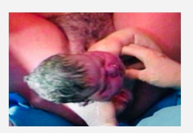
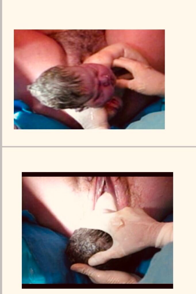
  • At this stage the head might start distending the perineum. The anus starts dilating and the head is seen at the vulva. It keeps receding between contractions.
  • When the head distends the perineum check if the perineum is stretching well.
  • Place the left hand on the advancing head with fingers spread equally over the vertex towards the bregma to stop any sudden explosive effort during and after crowning of the head. With the right hand guard the perineum, holding it with the pad.
  • Check if the perineum is stretching. If not, give an episiotomy at the height of a contraction if there is any indication that the head is about to crown.

Crowning of the Head

Next is the crowning of the head.

The parietal eminences pass through the bony outlet. At this stage the head no longer recedes between contractions.

Tell the mother to stop pushing as this might lead to a rapid delivery of the head and consequent brain damage. Ask her to pant. Research has shown that a series of short pushes are more effective than a long pus.

It is really hard work! So keep encouraging the mother with kind words and warmth.

Extension of the head

Assist the extension by grasping the parietal eminences with your left hand. Let the head come out slowly and naturally. Feel for the cord around the baby’s neck. If it is there, slip it from the baby’s neck over the head. If it is too tight, place two artery forceps on the cord and cut it between them. When the nose and mouth come out, wipe away the mucus with a sterile swab.

By this point the whole head should be out. The head will have restituted and rotated spontaneously to face the mother’s left or right thigh. 

This shows you that the shoulders have descended and rotated to the anterior posterior diameter.

Delivering the Shoulders by Lateral Flexion of the Body

Place one hand above and one below the fontal head. Depress the head gently toward the anus/neck, making sure it is neither twisted nor bent sideways till the anterior shoulder is free.

The delivering of anterior shoulder

Remind your assistant to give syntometrine intramuscularly (in a single dose). Guide the head upwards in the direction of the mother’s abdomen.

The delivering of posterior shoulder

The posterior shoulder will escape smoothly over the perineum. The rest of the body will be born by lateral flexion. Ask your assistant for the time and note the time of birth. Place the baby at a slight slant to drain the mucous. Put the baby on the baby towel, clamp and cut the cord. Give the APGAR score to the baby. Show the baby to the mother to identify the sex of the baby.

Ask your assistant to continue with the immediate care of the baby. Continue with the delivery of the placenta by using control cord traction. Check the placenta for completeness and/or malformation. Measure blood loss.

Do the first examination of the baby. Weigh the baby. Do a post natal examination and record all the findings. Give the mother a hot drink and transfer her to the postnatal ward.

Read More

Oyo State College Of Nursing And Midwifery Begins Sale Of 2024/2025 Admission Forms

The Oyo State College of Nursing and Midwifery (OYSCONME) has commenced the sale of Post UTME forms to prospective students aspiring to pursue the Basic Nursing Programme.

This was made known in a statement released by the Provost of the College, Dr Gbonjubola Owolabi. The Provost stated that the sale of forms will run from May 20th to August 16th, while the Post UTME examination is scheduled to be held on 26th-27th August after which the interview will follow on the 2nd to 6th September 2024.

She reiterated that the public should not provide personal or financial information to any unofficial source claiming to be associated with the College because she would never request for sensitive information through unofficial channels.

Dr. Owolabi charged interested candidates to follow the process of online application through the College website www.oysconme.edu.ng or contact the ICT Center of the College for further enquiry. She added that the College will not compromise her standards as admission is strictly on merit and the form is meant for those who have attained the cut-off mark of 200 and above.

The Provost further advised that payment should not be made through an Individual/Agent or Personal Bank account details asides from the link www.oysconme.edu.ng through which application form fee is expected to be made.

She noted that students would be awarded the academic certificate of National Diploma upon completion of the program.

Plateau State College Of Nursing And Midwifery, 2024 JAMB Cut-Off Mark

Plateau State College of Nursing Sciences, Vom announces the 2024 Jamb Cut-Off Mark for admission into the 2024/2025 Academic Session.

Having been included in the JAMB portal, the Plateau State College of Nursing Sciences is anticipating to commence academic activities in the 2024/2025 session. Consequently, at the Management meeting of 13/05/2024, a cut-off mark of 170 was agreed for students who sat for JAMB in the 2024/2025 academic session.

This is for your information, please.

Job Alert: State Technical Malaria Lead at the Management Sciences for Health (MSH) – 9 Openings

0

The Management Sciences for Health (MSH), a global health nonprofit organization, uses proven approaches developed over 40 years to help leaders, health managers, and communities in developing nations build stronger health systems for greater health impact. We work to save lives by closing the gap between knowledge and action in public health.

We are recruiting to fill the position below:

Job Title: State Technical Malaria Lead

Job Requisition ID: R4064
Locations: Bauchi, Sokoto, and Kebbi
Job type: Full-time
Department: Malaria Program
Slot: 9 Openings (3 per State)

Main Purpose of the Job

  • The State Technical Malaria Lead will be responsible for supporting the State Coordinator with technical leadership of the program by providing malaria technical expertise in the conceptualization and implementation of state systems and processes to support effective and efficient delivery of malaria services.
  • S/he will serve as a resource for up-to-date technical information on malaria control and elimination to USAID PMI and key stakeholders including the State Malaria Elimination Program.
  • The position will oversee malaria services quality assurance initiatives at the state level including improvements to diagnosis through RDTs and microscopy, and accurate treatment of both severe and uncomplicated cases of malaria using national treatment guidelines; IPTp, iCCM, and SMC implementation where applicable.

Overview

  • The Presidents Malaria Initiative for States (PMI-S) is a five-year PMI/USAID flagship malaria project implemented through a consortium led by the Management Sciences for Health (MSH).
  • The project is supporting the Government of Nigeria through its agencies at the federal, state, Local Government (LGA) and community levels to reduce under-five and maternal mortality by delivering quality services for management of malaria, its complications, and prevention.
  • PMI-S project focuses on strengthening the capacity of the National Malaria Elimination Program (NMEP) and State Malaria Elimination Program (SMEPs) for the implementation of the National Malaria Strategic Plan 2014-2020.

Specific Responsibilities

  • Provide technical leadership in the development and monitoring of the state malaria annual work plan (AOP) and work closely with the State Coordinator to implement and monitor departmental work plans.
  • Engage the State Malaria Elimination Program, SPHCDA, HMB, and relevant SMOH entities and partners to share progress, accomplishments, and challenges and ensure common understanding of current as well as future malaria technical direction.
  • Interacts with the key stakeholders in the malaria partnership at state level
  • Collaborate with the State Coordinator and the Finance and Operations Manager to optimize and utilize project resources in the most efficient way to achieve project results.
  • Lead and supervise the project’s malaria technical team at the state level
  • Build capacity of SMEP and roll out a mentoring and supportive supervision program at all levels of the state health system
  • Provide technical contributions to the state M&E/HSS officer in the development and implementation of a project surveillance monitoring and evaluation (SME) system including tracking the malaria cascade (persons with fever, tested with RDT/microscopy, positives treated with ACT, and outcome of treatment) and the related commodity data.
  • Provide TA to the SMEP on effective engagement of private sector health service providers in planning and implementation of state malaria elimination programs
  • Contribute to writing project reports, documentation of good practices, and technical publications
  • Contribute to visibility of PMI for States Project work through innovative presentation of project approaches and results at state, federal and international levels

Qualifications and Experience

  • Advanced Degree in Health and Postgraduate qualification in Public Health or related discipline
  • Should have good understanding of the Nigerian health system and the inter­relationships within the public and private health sector
  • Knowledge and minimum of three years of progressively responsible experience working on malaria control in public and private in Nigeria
  • In depth knowledge of malaria and public health principles with proven technical skills in malaria, including malaria case management, and integrated community case management of childhood illnesses
  • Experience working with Ministry of Health and other Health Departments/Agencies on policy and strategy formulation at national and/or subnational levels;
  • Experience with broader human resource capacity building
  • Nigerian with good understanding of local context.
  • Significant experience in project management, program coordination and sound negotiation skills with malaria partners;
  • Excellent writing, communication and presentation skills.

Application Closing Date
Not Specified.

How to Apply
Interested and qualified candidates should:

Click here to apply

Management Of Normal Labour And Vaginal Examination In Labour For Midwives

Physiological Changes in the First Stage of Labour

This is the stage of dilatation of the cervix.

On average, it lasts eight to twelve hours in a primigravida and six to eight hours in a multipara.

It should not go beyond 14 hours in either.

This stage is characterized by the uterus doing an immense amount of muscular work in the form of contracting and relaxing.

Contractions are involuntary in that they do not come on through voluntary effort of the woman nor can they be voluntarily stopped. 

They are peristaltic and regular.

Contractions at the start of labour come every 10 to 15 minutes and last between 30 to 45 seconds. 

They increase in frequency and strength as labour progresses until they are separated by a minute or two.

Towards the end of the first stage they last for one minute. The relaxation phase during which the muscles remain retracted also shortens.

Each contraction begins with a gradual build-up towards a peak of intensity. 

This is followed by a relaxation phase.

During the relaxation phase, the muscle recovers and gets ready for the next contraction.

This relaxation phase is important to both the foetus and the mother during the first and second stages of labour.

During a contraction the circulation through the uterine wall is reduced and the foetal heart rate is slowed but regains its normal rate as soon as the contraction has passed.

If there is increasing foetal tachycardia between contractions or if the bradycardia is prolonged after each contraction, then foetaldistress sets in. If the uterus contracts continuously the foetus dies from lack of oxygen (anoxia).

The mother is also able to relax during the relaxation phase.

However, if the uterus contracts continuously, the mother also gets very exhausted because the uterus uses up a lot of energy during contractions.

If this goes on for too long, as is the case during prolonged labour, her energy stores become depleted and maternal distress sets in.

As the mother will not be able to eat or absorb much by mouth, she should be given supplementary carbohydrates intravenously. 

In a normal first stage, oral fluids to which additional sugar has been added are sufficient.

As the uterus contracts and retracts more and more, the upper muscular part becomes progressively thicker.

The less muscular lower segment is pulled upwards over the presenting part and becomes thinner.

The cervix becomes effaced. 

The effacement is followed or accompanied by progressive dilation of the cervix until full dilatation when the uterus becomes a continuous cavity with the vagina.

A fully dilated cervix is 10 cm dilated. During the first stage the uterine cavity gets progressively smaller but the foetus moves down very little.

The cervix becomes effaced. The effacement is followed or accompanied by progressive dilation of the cervix until full dilatation when the uterus becomes a continuous cavity with the vagina.

A fully dilated cervix is 10 cm dilated. During the first stage the uterine cavity gets progressively smaller but the foetus moves down very little.

A fully dilated cervix is 10 cm dilated. During the first stage the uterine cavity gets progressively smaller but the foetus moves down very little.

The presenting part helps in dilating the cervix.

During this stage the woman should not use her voluntary efforts to bear down as this will exhaust her unnecessarily and may cause oedema of the cervix and/or foetal distress.

Management of Normal Labour in the
Admission Room

The proper management of labour is essential, if you are to avoid problems or to detect them early when they occur.

The patient will come to you believing she is in labor. You should be able to assess and decide whether she is in labour or not.

The patient may be in early labor, but often she might arrive in the late second or even third stage.

If you are sure she is not in labour send her home to wait. If she is in labour, keep her in the ward and continue monitoring her progress.

Danger, especially to the fetus, can arise suddenly and unexpectedly.

No labor should be assumed normal until the fourth stage has successfully concluded.

Admission

You would take the patient’s personal history, conduct a physical examination and carry out tests/investigations.

On admission, you should check the woman’s antenatal card for any identified risk factors.

It will also help you to see if there were any abnormalities during her pregnancy.

The records will also have information on her medical and obstetric history. If she has not been attending an antenatal.

Once this information has been established, find out more about the present labor.

Take her history, do an examination, and carry out the necessary investigations to establish the stage of labor and the state of the mother and the fetus.

History Taking

A detailed personal history should have been taken during pre-natal care.

However, if this has not been done, this is a good time to get it recorded.

Make sure the names are correctly spelled because this can eventually result in problems when registering the baby.

Review the last day of menstruation to calculate the expected date of delivery.

Check her age, parity, and contraceptive

Assuming that a detailed personal history had been taken during pre-natal care, you should now take information about the following:

➢ Any presence of show

➢Presence or absence of contractions

➢ Onset of contractions and their characteristics

➢ Activity of the foetus

➢ Rupture of the membranes

➢ Any treatment given

Head-to-Toe Physical Examination

Conduct an abdominal examination checking for:

➢Height of fundus

➢ Over-distension of the abdomen, scars or other abnormality

➢Over-distension of bladder

➢ Possible presence of twins or multiple pregnancy

➢ Contractions – frequency, length, type etc

You should also check on the presentation.

Which part of the foetus is at the pelvic brim?

Is it a head (cephalic) or the buttocks (breech)? Check the attitude; whether the head is well flexed or extended.

A well-flexed head presents the smallest diameter and delivers easily.

A deflexed head presents a larger diameter and causes delayed or obstructed labour.

Finally check the position of the relation of the foetal parts to the mother.

This is confirmed through a vaginal examination by checking the position of the foetal occiput relative to the mother.

The position of the foetal spine is the same as that of the occiput.

Vaginal Examination in Labour

This is an important examination as it can give you a lot of information, which you might not get from an abdominal examination.

On the other hand, if you do it often it is uncomfortable for the woman and you might introduce an infection into the uterine cavity, especially if the membranes have ruptured.

To avoid infections, you should scrub and put on gloves as you would for any other sterile procedure.

Then thoroughly swab the perineum of the woman with an antiseptic solution such as Savlon or Hibitane, or boiled water if these are not available.

A vaginal examination is necessary to:

Check if the patient is in labour and what stage of labour (this is done on admission of the patient)

Assess the progress of labour (this is done every four hours during the first stage of labour)

The degree of effacement and dilatation of the cervix and the station of the head relative to the ischial spines will give you the necessary information.

Remember: Do not do a vaginal examination if the mother has an antepartum hemorrhage, because if there is placenta praevia, severe hemorrhage will occur.

A vaginal examination is contraindicated if the mother has ante-partum haemorrhage unless it is performed in an operating theatre. In the case of pre-eclampsia, the procedure should be performed only after giving a sedative.

Vaginal Examination

Arrange your vaginal examination pack with cheatle forceps and pour solution.

Scrub your hands for at least five minutes.

Glove yourself methodically to prevent contamination.

Explain the semi-lithotomy position that should be maintained during the examination to the mother.

Swab the vulva and drop the swab methodically (used swabs should be decontaminated in jik before disposal).

Ask the mother to breathe in and out while you perform digital examination.

With the right hand, gently insert the fingers obliquely inside the vagina with the thumb, facing the symphysis pubis. Your left hand.

The fingers are directed along the anterior wall of the vagina. The wall should feel soft and dilatable while the vagina should be warm and moist.

The fingers are then directed upwards to the position of the cervical Os.

At times the Os is not felt readily, the fingers should then be directed backwards and upwards.

While performing a vaginal examination, you should observe the mother’s non-verbal communication.

The Vagina

The Cervix

➢ Is it bruised or oedematous?

➢Is it firm or soft?

➢ Is it taking up, that is effaced?

➢ How much is the Os dilated?

Do not insert more than two fingers in the cervical Os.

The Membranes

After deciding the state of the cervical Os, check for presence of membranes. Note the following:

Are they ruptured or intact?

If intact are they bulging?

The Cord

Is it presenting or prolapsed?

If prolapsed is it pulsating?

The Presenting Part

Next, determine the level of the presenting part. You should ask the following questions:

Is it fitting the pelvis and cervix well?

If it is the head, can you feel a suture or fontanel? Which one?

Is it well flexed?

If the head is at the brim it, will not be felt vaginally unless you push it down with your left hand which is on the mother’s abdomen

If the head has just engaged, it can be touched or just be tipped

If the head is deeply engaged, the head is felt at the level of the ischial spines.

While performing a vaginal examination, you should observe the mother’s non-verbal communication.

The Presenting Part

The station or level of the presenting part is the level to which the presenting part has descended in the pelvis.

The level of the presenting part is expressed in relation to the easily palpable ischial spines.

You can also state if it is above the brim, at the brim, in the cavity or at the outlet.

Remember that if the presenting part is at the brim then it is at station -5.

As the head descends down in to the cavity of the pelvis it decreases from -5, -4, -3, -2, -1. By the time it is at the ischial spines, it is said to be at ‘zero station’. It reappears from the pelvic outlet into the perineum, which is classified as +1, +2, +3, and +4.

By the time it is seen at the perineum it is at

To get the position right, you have to palpate the sutures and fontanel to determine their position relative to the pelvis. 

This can be confirmed by abdominal examination.

In a breech or a face presentation, the reference points on the foetus are the sacrum and the chin or mentum respectively. A soft mass is felt.

Foetal genital parts or the anus may be felt with the examining finger. 

The position is determined by the position of the sacrum in relation to the side of the mother’s pelvis. The left side is expressed as left sacro-anterior (LSA) or the right side, which is known as right sacro-anterior (RSA).

In a vertex presentation, when the occiput is persistently posterior, this causes prolonged labour.

In a cephalic presentation, you will feel the hard head sutures and fontanel.

Determine whether it is the anterior or posterior fontanel by its shape.

If it is the posterior fontanel, then the position is occipito-anterior.

If it is the anterior fontanel then the position is occipital-posterior.

Face presentation is important as the chin is the facial landmark. 

Note how the position of the chin is related to the mother’s right thigh (RT) or left thigh (LT), side of the pelvis and the anterior and posterior position expressed as RMP (right mentoposterior). You should also note the brow or sinciputpresentation on vaginal examination (VE).

You should be able to feel the brow, frontal sutures and possibly the posterior fontanel. The extension is usually a temporary presentation, which converts to face or occiputpresentation during labour.

However, at times it persists and in such instances, a caesarean section is usually called for.

Note whether the long axis of the body of the foetus is perpendicular to that of the body of the mother.

A left acromiodorso posterior (LADP) indicates that the baby’s lower shoulder is to the mother’s left and its back is towards her back.

A compound presentation is when the hand or occasionally a foot lies alongside the head.

The Pelvis

Next, check for moulding or caput succedaneum.

What is moulding?

Moulding is when the diameters of the foetalskull are reduced in size. During labour the bones of the foetal skull tend to overlap at the sutures so that the head can easily pass through the birth canal.

During a vaginal examination this is how you should check for moulding:

In cephalic presentation, run the finger on the head feeling for the sutures

Judge the degree of moulding by feeling the amount of overlapping of skull bones

Check for caput.

The pelvis is assessed to check if it is adequate

The following factors should be checked:

  • Is it roomy?
  • Are the sidewalls well spaced?
  • Can you touch the promontory of the sacrum easily?
  • Is the pubic arch wide enough?
  • After checking for moulding, direct the fingers behind the head and make an effort to reach the sacro-promontory. The palm of the hand.

With the palm facing downwards run the two fingers down along the hollow of the sacrum and determine the shape. The hollow of the sacrum should be curved.

The two fingers are then moved to where the ischial spines are located on either side of the pelvis. Run the finger along this area to determine whether or not the ischial spines are unduly prominent. Or else you can stretch your fingers to see if the spines are prominent. The ischial spines should not be prominent.

As you move the fingers with the palm facing upward, on reaching the pubic arch check if it can accommodate two fingers. The apex usually should accommodate two fingers.

Make a fist facing downwards then place the fist between the ischial tuberosities.

The Intertuberosity should accommodate four knuckles.

The Discharge

Withdraw the fingers and check if there is:

➢Any vaginal discharge

➢Any smell

➢Any liquor or meconium staining

➢Any bleeding

The following steps should be taken as part of your investigation:

  • Take a urine sample for albumin and sugar
  • Check for acetone, especially if the patient is in prolonged labour
  • Take blood for haemoglobin and cross matching if the patient is anaemic or might need an operation

By this time you will have gathered enough information as to the stage of labour and whether the patient belongs to the ‘at risk’ category and needs referral or not.

Read more

Normal Pregnancy: Labour, Puerperium, And Stages of Labour In Pregnancy

Labour

Labour is described as the process whereby the foetus, placenta and membranes are expelled through the birth canal after 28 weeks of gestation.

Labour, can be either normal or abnormal.

Normal labour has several important characteristics, which you should always remember.

These are:

Duration – completed within 18 hours

  • Occurs at term between 38 and 40 weeks
  • Is spontaneous
  • The foetus presents by the vertex
  • Has no complications to either mother or baby
  • The newborn child requires minimal or no

The Onset of Labour

  • A midwife should ensure women have sufficient information to assist them to recognize the onset of true labour.
  • A pregnant woman would be best placed to diagnose the onset of labour herself.
  • Some young women, especially the primigravidae (those pregnant for the first time), may fail to recognize true labour.
  • It is important that you help them differentiate between false and true labour signs.
  • The contractions of true labour are regular and intense.
  • In false labour, the contractions are sporadic (Braxton-Hickscontractions).
  • False contractions occur during the last weeks of pregnancy.

Signs and symptoms that gives evidence that the mother is in labour:

  • Contractions of the uterus, which are increasingly strong, painful and regular.
  • The cervix is taken up into the lower uterine segment causing dilatation of the cervix.
  • There is a mucoid blood stained discharge, which is called show.
  • Sometimes there is rupture of membranes with drainage of liquor amnii (amniotic fluid).
  • The causes that trigger the onset of labour are not known.
  • However, many theories have been offered which indicate that both hormonal and mechanical factors play a big part.

Hormonal Factors

It is believed that close to term progesterone levels in the body fall, while at the same time levels of oestrogen (which is responsible for sensitizing the uterine muscles) rise.

The fall in progesterone levels is important because it has effect on muscle contractions.

The rise in oestrogen levels meanwhile triggers the release of oxytocin, which causes uterine contractions.

The foetal hypothalamus is believed to produce releasing factors, which stimulate the anterior pituitary gland to produce adrenocorticotrophichormone (ACTH).

Mechanical Factors

Uterine activity can also result from the mechanical stimulation of the uterus and cervix.

This may be due to over stretching, as in the case of multiple pregnancy and polyhydromnios, or pressure from the presenting part, when it is well applied to the cervix.

It appears that there is a combination of hormonal (from both mother and foetus) and mechanical factors that set labour in motion.

Pre-Labour or Premonitory Signs of Labour

This is the period two to three weeks prior to the onset of labour when a number of changes take place.

Lightening

Two to three weeks before labour, the lower uterine segment expands allowing the foetalhead to sink deep.

The descent of the head and the body of the baby gives space to the lungs, heart and stomach, which enables these organs to function easily.

The symphysis pubis widens and the pelvic floor softens and becomes more relaxed, allowing further descent of the uterus into the pelvis.

Frequency of Micturition

The descent of the foetal head increases pressure within the pelvis.

This limits the capacity of the bladder, which can cause irritation. 

The laxity of the pelvic floor muscles gives rise to poor sphincter control causing a degree of stress incontinence.

This pressure results in the congestion of circulation to the lower limbs.

Additionally the relaxation of the pelvic joint may give rise to backache.

Taking up of Cervix

The cervix is taken up gradually and merges into the lower uterine segment. Shortening of cervix is looked for when labour needs to be induced.

Contractions

The contractions of the uterus are coordinated by two pacemakers in the region of the cornua.

These are located where the fallopian tubes join the uterine body.

The muscle contractions start at the top corner of the uterus, spread to the fundus, and then downward.

During normal pregnancy, the uterus contracts intermittently but the contractions are not strong enough to overcome the resistance of a normal cervix and do not lead to its dilation.

The contractions of pregnancy become more frequent towards full term and get more painful and noticeable.

A multipara may have such ‘false pains’ for some days before the onset of true labour. They may come to hospital too early thinking they are in established labour. This is what is referred to as ‘false labour’.

Uterine Action

By the end of pregnancy the uterus is divided into two anatomically distinct segments, known as the upper and the lower uterine segments.

The upper uterine segment is a thick muscular, contractile area from where the contractions begin.

The longitudinal fibers retract, pulling on the lower segment and causing it to stretch, pushing the head down.

The lower uterine segment is thinner and develops from the isthmus of the uterus about eight to ten centimeters in length and is prepared for distension and/or dilatation. 

The lower segment stretches when being pulled by the longitudinal fibres. The force applied by the descending head or breech also aids the stretching.

The retraction ring which is an imaginary ridge, forms between the upper and the lower uterine segment.

It is present in every labour and is perfectly normal as long as it is not marked enough to be visible above the symphysis pubis.

Fundal Dominance

During a contraction the uterus feels hard to touch. At the beginning of the process, contractions are painless and involuntary, and are controlled by the nervous system under the influence of endocrine hormones.

The contraction starts at the upper part of fundus, spreading across, and by the time they reach the lower fundus, they last longer and are very intense.

The peak of the contraction is reached simultaneously over the whole uterus and fades from all parts together. This pattern allows the cervix to dilate and the contracting fundus to expel the foetus.

Polarity

Polarity describes the neuromuscular harmony between the two poles or segments of the uterus throughout labour.

The upper pole contracts strongly and retracts to expel the foetus.

The lower pole contracts slightly and dilates to allow expulsion of the foetus to take place.

Contraction and Retraction

When labour starts, approximately 280 days from the first day of the last menstrual period, the contractions change in character. They become regular and more painful.

Labour contractions differ from those of pregnancy in that they are followed by retraction.

This is characteristic of uterine muscle in labour.

The contracted muscle does not return to its original length when the contraction passes off.

Each succeeding contraction leads to further shortening of the muscle fibres so that the uterine cavity becomes smaller and smaller.

When talking about contractions, you as a midwife are concerned with three factors, namely the strength, the duration and the frequency of the contraction.

When you talk of the strength of a contraction, you identify it as one of three categories: weak, fair or fairly strong, and strong. 

The strength of a contraction is measured according to the time it has taken.

Thus, a contraction which takes 30 seconds is said to be weak, one that takes 30 to 40 is said to be fair or fairly strong and one that lasts for 40 to 60 seconds is said to be strong.

The duration refers to the time taken by a contraction, for example a weak contraction lasts for 10 to 30 seconds.

Frequency, on the other hand refers to the number of intervals between one contraction and the next.

If a mother has one contraction after every 45 minutes, the frequency is written as 1:45.

The Shortening and Dilation of The Cervix

Before labour begins, the cervix of a primagravida is a thick hard cone which protrudes into the vagina.

The canal is at least one inch long.

When labour begins the strongly contracting upper segment of the uterus starts retracting and getting shorter, while the thinner lower segment of the uterus gets pulled away from the presenting part. This stretches the lower segment.

The latter, in turn pulls the internal Os. This dragging away of the internal Os from the presenting part starts dilating the upper part of the cervical canal. 

This goes on until the canal is shorter and shorter and finally there is no canal at all.

The canal becomes part of the uterine cavity with only the undilated external Os and the thinly stretched cervix separating this cavity from the vagina. When this happens, we say the cervix has been ‘effaced’ or ‘taken up’ 

In a primigravida the cervix usually becomes almost fully effaced before any dilation takes place, while in multiparous women the two processes take place together. 

The cervix of a multipara might be already effaced and be dilated enough to admit a finger internal Os even before labour starts.

These signs of labour are assessed by doing a vaginal examination, though with experience, you can also get a good idea by doing a rectal examination.

As a midwife, the way you guide a mother has a great influence on the progress of her labour.

The Show

Throughout pregnancy the cervical canal is sealed by a plug of mucus known as an operculum. Together with the intact membranes this prevents organisms ascending into the uterine cavity.

When labour starts, the internal Os is pulled away from the foetal membranes and the canal is opened up. This releases the mucous plug which oozes out of the vagina mixed with a little blood. This is called the ‘show’.

The Stages of Labour

Labour is divided into four stages, although in real practice, the process is a continuous one and change from one stage to the other may not be clearly obvious. The four stages of labour are:

➢First stage,

➢ Second stage

➢Third stage

➢Fourth stage

The First Stage Of Labour

This is known as the stage of cervical dilatation.

This stage begins when regular, painful uterine contractions start and is detected clinically by the thinning and effacement of the cervix, followed by its dilatation.

The normally thick cervix becomes thinned out and stretched over the presenting part.

The first stage is completed when the cervix is fully dilated and the presenting part starts being expelled.

This stage has two phases, known as the latent and the active phase.

The contractions of the uterus dilate the cervix. 

The dilatation of the internal Os causes the separation of the chorion from the deciduaclosest to it.

A small bag of membranes is formed and is forced into the internal os by the intrauterine pressure.

At the beginning of each contraction, a little more amniotic fluid is forced into the bag of membranes. The head of the foetus then comes down like a ball valve and separates the amniotic fluid above it from that in the bag. 

The bag of membranes may remain intact until nearly the end of the first stage.

However, even if the membranes rupture early the cervix will still become dilated as it is drawn up over the presenting part by the retraction of the upper segment.

During the first stage, the foetus does not move downwards to any great degree. When a certain amount of fluid has left the uterus after the membranes have ruptured, a new form of pressure comes into play, namely foetal axis pressure. The upper pole of the fetus.

The upper pole of the foetus, in the breech, is pressed on by the fundus of the uterus, while the lower pole is pressed down onto the lower segment and cervix.

Should the membranes rupture early, foetalaxis pressure will operate at an early stage.

In modern practice, the membranes are often deliberately ruptured during labour because this is believed to encourage more efficient uterine action and shorten labour.

You should be cautious carrying out this procedure in this era of HIV.

The duration of the latent phase of labour need not be defined too accurately.

Dilation of the cervix from 0 to 3cm can take six hours, but slower progress may be normal and is perfectly acceptable provided that the woman is comfortable and in no way distressed.

Between 3 and 10cm dilatation (that is in the active phase of labour) the cervix should dilate at a rate of about 1 cm per hour, giving a theoretical duration of seven hours for this phase of labour in both primiparous and multiparous women.

During the early part of the latent phase the pains may not be very severe but towards the end of the active phase they are often very distressing, constituting the most painful part of labour.

Vomiting and reflex shivering are common at the end of the active phase of the first stage of labour.

By now the membranes have ruptured. If the membranes remain intact when the cervix is fully dilated, the onset of the expulsive stage may be delayed.

This is because the cervix does not receive the pressure of the head, which helps to stimulate the uterus to increase its activity.

If the membranes remain intact after full dilatation, they should be ruptured with toothed forceps or a sterile plastic amnihookduring a contraction.

Second Stage Of Labour

This starts with the full dilatation of the cervix and the expulsion of the presenting part and finishes with the complete delivery of the baby.

As you know, there may be very little descent of the foetus during the first stage. However, in the second stage, the resistance offered by the lower uterine segment and the cervix has been overcome and the presenting part can be pushed down onto the pelvic floor.

The resistance of the pelvic floor then has to be overcome by uterine contractions, aided by the action of the voluntary muscles of the abdominal wall and the diaphragm.

In the absence of an effective epidural block, full dilatation of the cervix is accompanied by a bearing down sensation during contractions and women are usually encouraged to push.

As the contraction comes on, the woman takes a deep breath, then holds it and subsequently bears down with all the force of her abdominal.

These partly voluntary, partly reflex expulsive efforts place the foetus under additional stress and pushing should, therefore, not be allowed to continue for more than one hour. 

If delivery is not imminent, assistance in the form of forceps delivery or even a Caesarean section may be necessary.

Third Stage Of Labour

This stage commences immediately after the birth of the baby. It includes the delivery of the placenta and membranes as well as the control of bleeding.

At this stage the uterus contracts down to follow the body of the foetus as it is being born. As the cavity of the uterus becomes smaller, the area of the placental site is diminished.

The placenta is then cut off from the spongy layer of the decidua basalis. Further uterine contractions expel the placenta from the upper segment into the lower segment and through the vaginal vault.

This process, whereby the placenta leaves the upper segment to the lower segment and through the vagina, is referred to as separation and descent.

Fourth Stage Of Labour

This is the period from the delivery of placenta and membranes to the end of the first hour postpartum. The uterus is firm at level of two fingers breadth above the umbilicus. Restoration of physiological stability is established.

During this period myometrial contractions and retraction, accompanied by vessel thrombosis, operate effectively to control bleeding from the placenta site. 

Failure of this mechanism could result in excessive blood loss (postpartum haemorrhage(PPH)) that could be life threatening.

The mother should be closely observed for haemorrhage, urine retention or hypotension.

The mother and child relationship should be initiated and encouraged, as it has an effect to the subsequent quality of their relationship and bonding.

Read more

Bayelsa State College Of Nursing Sciences Begins Sale Of 2024/2025 Forms

This is to notify the general public that the last batch of entrance forms for registered Nurses/ registered Midwives is out from 1st June to 30th June, 2024.

Candidates are to pick their forms at the School registry with the proof of payment/ bank teller from the bank.

Note: This is not for the ND/HND program that requires Jamb results.

Admission Requirements

Interested applicants must possess five (5) credit passes in English Language, Mathematics, Biology, Physics, and Chemistry in not more than two (2) sittings.

A combination of WAEC and NECO or GCE is allowed. Applicant must be less than 17 years of age.

Mode Of Payment

Amount to pay: Twenty thousand naira (20,000)

Account Number: 0097373620

Account Name: Bayelsa State College Of Nursing Science

Bank: Sterling bank

Date For Entrance Exams

Thursday 4th to Saturday 6th July, 2024

Mode Of Examination

Computer Based Test (CBT)

Venue For Examinations

ICT Center, BYSCON, and the time is 9 am prompt.

For more information, call: 08138661996